3-D Imaging Can Make Breast Cancer Treatment More Effective
Researchers say newer MRI tools can reveal tumors missed by standard screening methods. It can also provide a better understanding of treatment.
Posted — UpdatedIn the fall of 2005, Marlyn Smith had what she thought was an infected bug bite on her right breast. She played it safe and had it checked.
"We did a mammogram, ultrasound and they both came back negative," Smith said.
Still, the condition grew worse. Smith had inflammatory breast cancer, a rare but aggressive form of the dieases often missed by standard screening methods. She did not know it was cancer until three months later when she came to Wake Radiology for an MRI.
"It was overwhelming. When I first saw it, it showed all the cancer," Smith said.
Dr. Glenn Coates said an MRI is too expensive to replace mammograms and ultrasound and he said they are still effective tools for early detection, but a recent study in the New England Journal of Medicine shows that for women who have a positive needle biopsy, more detailed imaging is needed.
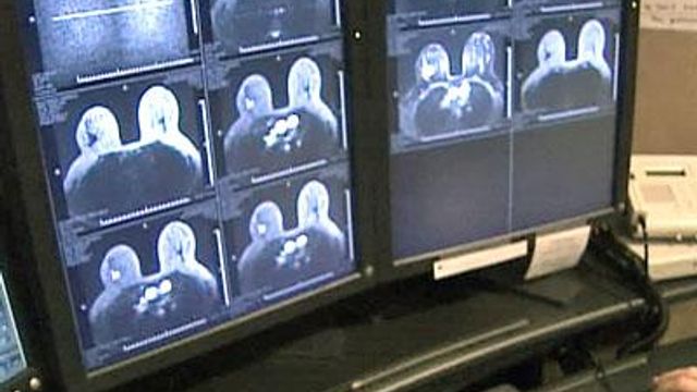
In about 1 out of 10 cases where standard screening diagnosed cancer in one breast, MRI found early stage cancer in the other. The MRI equipment at Wake Radiology has other advantages. They're one of the few centers in the state able to look at the breast scans in three dimensions.
From the flat two-dimensional view, one tumor measured two centimeters. However, that measurement is true only from one perspective. A three-dimensional image showed another view.
"The tumor is actually 5-1/2 centimeters in size," Coates said.
The 3-D view also reveals several satellite tumors hidden in the previous 2-D image. Coates said radiologists can correctly interpret a tumor's true size through two dimensional images, as they move through the different "slices" of images provided by MRI.
A better understanding of the staging of the disease also helps determine treatment.
Marlyn Smith had surgery to remove the breast and several lymph nodes. She had radiation and is near the end of her schedule of chemotherapy treatments. She said when the symptoms first appeared, she knew something was wrong, even though the early tests were negative. Other women, she said, should trust their instincts and get a second or third opinion.
Most insurance providers will cover MRI costs if there is a positive needle biopsy. Most will also cover an MRI test, even if standard screening is negative, but the symptoms concern the patient and their doctor.
• Credits
Copyright 2024 by Capitol Broadcasting Company. All rights reserved. This material may not be published, broadcast, rewritten or redistributed.


